Peritonsilar Abscess Drainage
From Guide to YKHC Medical Practices
written by Dr. Travis Nelson
Peritonsillar abscess is an uncommon but not rare complication of bacterial pharyngitis and tonsillitis. It is distinguished from peritonsillar cellulitis by the presence of soft palate swelling and/or uvular deviation. If swelling is severe the airway can become impaired, thus peritonsillar abscess drainage is a vital skill. CT is not necessary to diagnose peritonsillar abscess though may be indicated to rule out deep space infection. Bedside US using a sheathed endocavitary (vaginal) probe may be used to assist in abscess localization, though is sometimes limited by trismus.
Technique
- Due to the proximity of major vessels of the neck and the lack of vascular surgery at this facility, scalpel I&D of peritonsillar abscesses is not recommended at YKHC.
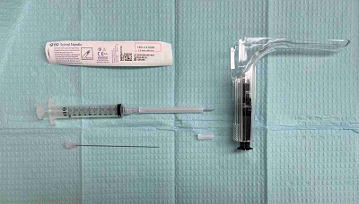
- Needle drainage is performed as follows (see supplies setup in image):
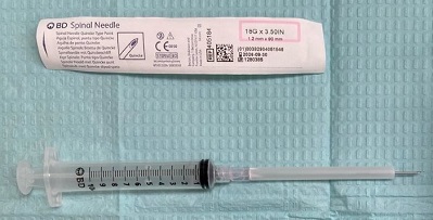
- An 18 gauge 3 ½ inch spinal needle is prepared by removal of the trochar and cutting off 1 cm from the plastic sheath surrounding the needle.
- Attach prepared 18 gauge needle to a 10-20 cc syringe.
- Ensure suction is available for bleeding, often the patient is allowed to hold suction themselves.
- Although visualization via tongue blade or laryngoscope is possible, the easiest method for visualization I’ve found is to remove the top of a disposable speculum and insert it into the oropharynx while placing downward pressure to move the tongue out of view. The speculum has its own light source, negating the need for a headlamp or assistant holding a light.
- Topical benzocaine (Hurricaine) spray has somewhat fallen out of favor due to r/o methemoglobinemia though is still frequently utilized for anesthesia.
- Once visualization is achieved, the needle is inserted into the abscess pocket while drawing back on the syringe. Typically it is inserted at the superior pole, then if not successful reattempted at the middle pole, then if still not successful reattempted at the inferior pole.
- Send any purulent fluid drained for culture to guide antibiotic management.
- There is typically scant bleeding following the procedure that resolves without intervention. Any persistent bleeding or concern for vascular trauma should be discussed immediately with ENT and vascular surgery.
Resources/References
- Galioto NJ. Peritonsillar Abscess. Am Fam Physician. 2017 Apr 15;95(8):501-506. PMID: 28409615.
- Peritonsillar Abscess (YKHC Common/Unique Diagnoses)
- Peritonsillar Abscess Evaluation & Treatment YKHC Clinical Guideline

