Central Line Insertion
Central venous catheter insertion is indicated for access, hemodynamic instability and medication administration. Although pressors may be run through peripheral veins this is not optimal and initiation of pressors should be followed shortly by central line placement. Common complications of central line placement include failure of procedure, cannulation of artery, pneumothorax, bleeding and line infection.
Access can be achieved via internal jugular (IJ), subclavian or femoral vein catheterization. US guided IJ catheter placement is the optimal approach for most patients. The subclavian vein cannot be viewed well on US due to distortion from the clavicle, increasing risk of iatrogenic pneumothorax. The femoral vein may be rapidly and safely accessed without use of US and femoral line placement may be preferred when there is limited time for US guided placement. These lines are prone to infection however and are typically replaced by IJ catheters once the patient is stable.
Technique
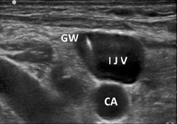
US guided IJV insertion (can be applied to femoral vein): Strict use of sterile technique throughout procedure is essential to preventing line infection. Provider should be sterilely gowned and gloved and patient should be sterilely draped. If patient can tolerate it, placement of patient in slight Trendelenburg position allows increased blood flow to the IJV. Prior to procedure flush the catheter with normal saline, cap the ends of the catheter and pull back (‘load’) the guidewire into its sheath. Cleanse skin with chlorhexidine and drape patient. Place vascular US probe into sterile sheath. On US identify the vein and distinguish it from the carotid artery:
- vein will be fully compressible compared to carotid artery
- vein will not have pulsatile flow on auditory Doppler
- vein will not have pulsatile color flow. Once the vein is identified inject a small amount of lidocaine subcutaneously above anticipated insertion site.
In our current central line kits, there are two options for initial needle placement:
- 18 gauge straight needle
- 18 gauge needle encased in soft catheter. Either of these may be used with a normal syringe or with the introduction syringe (hollow syringe, negates need for syringe removal prior to placement of guidewire).
Under ultrasound guidance, locate IJ at point most separate from carotid artery. Measure depth to vein and place needle on skin at distance equal to depth and at 45-degree angle towards vein, which will ensure the tip of the needle is visible in the plane of the ultrasound at the point it enters the IJ. Insert slowly while withdrawing on syringe until blood return. If not using introduction syringe remove syringe and place guidewire into needle. Never let go of the guidewire. Once guidewire is threaded remove needle over guidewire. Prior to dilation place the ultrasound and ensure you can visualize the guidewire in the vein.
Make a small skin incision with #11 blade at needle insertion site and thread dilator over guidewire. Remove dilator and thread catheter over guidewire until guidewire comes out brown colored port. While holding guidewire continue threading the catheter to desired depth (typically 15 cm for R IJ, 18 cm for L IJ though this can be adjusted based on patient size and common sense). Remove guidewire in full. Flush all ports and secure line to skin with included suture material. Obtain a portable CXR to confirm correct placement and adjust depth as needed.
Tips
- if patient is severely volume depleted IJ may collapse completely during inspiration. It may be necessary to thread the guidewire between breaths only during expiration.
- often it is easy to get blood flow in needle / syringe but the guidewire fails to thread and meets resistance. This is typically because either the needle shifted position and is no longer in the vein or the patient’s anatomy is such that it is difficult to thread the guidewire. Once the guidewire is used, it often kinks. When the guidewire meets resistance, do not try to force the guidewire, as you risk creating a false track. Remove guidewire completely and see if there is still blood flow from needle. If there is no flow then the needle has shifted position. Remove, apply pressure until bleeding stops and repeat procedure. If there is still good flow from needle, then open another kit and try a different guidewire.
- if pulsatile flow from needle, STOP PROCEDURE, remove needle and place pressure on wound.
- if cannulation of artery occurs do not remove central line. Transfer patient for removal under vascular surgery.
Resources/References
- IJ : Ultrasound Guided Internal Jugular Vein Cannulation NEJM YouTube Video
- femoral : https://www.nejm.org/doi/full/10.1056/NEJMvcm0801942
